

Difference Between Animal and Plant Cells Under a Microscope
Plant and Animal Cell Differences
Contents
- The Differences Between Plant and Animal Cells
- Investigation: Examining Plant Cells Under the Microscope
- Aim
- Apparatus
- Method
- Investigation: Examining Animal Cells Under the Microscope
- Aim
- Apparatus
- Method
- Questions
Now that we have looked at the basic structures and functions of the organelles in a cell, you would have noticed that there are key differences between plant and animal cells. The table below summarises these differences.
| Animal Cells | Plant Cells |
| Do not contain plastids. | Almost all plants cells contain plastids such chloroplasts, chromoplasts and leucoplasts. |
| No cell wall. | Have a rigid cellulose cell wall in addition to the cell membrane. |
| Contain centrioles. | Do not contain centrioles. |
| Animals do not have plasmodesmata or pits. | Contain plasmodesmata and pits. |
| Few vacuoles (if any). | Large central vacuole filled with cell sap in mature cells. |
| Nucleus is generally found at the centre of the cytoplasm. | Nucleus is found near the edge of the cell. |
| No intercellular spaces found between the cells. | Large intercellular air spaces found between some cells. |
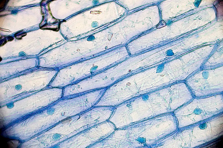
Aim
To study the microscopic structures of plant cells
Apparatus
- onion
- blade
- slides and coverslips
- brushes
- compound microscope
- tissue paper
- forceps
- dropper
- iodine solution
- watchglass
- petri dish containing water
- iodine solution
Method
- Peel off the outer most layer of an onion carefully, using a pair of forceps.
- Place the peeled layer in a watchglass containing water. Make certain that the onion peel does not roll or fold.
- Using a scalpel or a thin blade, cut a square piece of the onion peel (about 1 cm2).
- Remove the thin transparent skin from the inside curve of a small piece of raw onion and place it on a drop of iodine solution on a clean slide.
- Cover the peel with a coverslip ensuring that no bubbles are formed.
- Using a piece of tissue paper wipe off any excess iodine solution remaining on the slide.
- Observe the onion skin under low power of the microscope and then under high power.
- Draw a neat diagram of 5-10 cells of the typical cells you can see.
Aim
to study the microscopic structures of human cheek cells under a compound microscope
Apparatus
- clean ear bud
- clean slide
- methylene blue
- dropper
- water
- tissue paper
- forceps
- microscope
Method
- Place a drop of water on a clean glass slide.
- Using a clean ear bud, wipe the inside of your cheek. The ear bud will collect a moist film.
- Spread the moist film on a drop of water on a clean glass slide, creating a small smear on the slide.
- Use a coverslip to cover the slide gently.
- Place one or two drops of stain on the side of the cover slip.
- Use a piece of tissue to remove the excess dye.
- Observe the cheek cells under low power magnification and then under high power magnification.
Questions
- What are the shapes of epidermal cells of the onion peel and the human cheek cells?
- Why is iodine used to stain the onion peel?
- What is the difference between the arrangement of cells in onion cells and in human cheek cells?
- Why is a cell considered the structural and functional unit of living things?

[Attributions and Licenses]
Difference Between Animal and Plant Cells Under a Microscope
Source: https://nigerianscholars.com/tutorials/introducing-the-cell/plant-animal-cell-differences/
0 Response to "Difference Between Animal and Plant Cells Under a Microscope"
Post a Comment